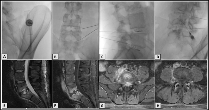
Figure 2.
Case 1. Procedural imaging and magnetic resonance imaging (MRI) 8 weeks postprocedure. (A) Fluoroscopy demonstrating bone marrow aspiration. (B) Anteroposterior view fluoroscopy demonstrating intradiscal procedure. (C) Lateral view fluoroscopy demonstrating intradiscal injection with discograms. (D) Contralateral oblique fluoroscopy demonstrating bone marrow concentrate (BMC) intra-articular facet injections at L3-4, L4-5, and L5-S1. (E) T2-weighted lumbar MRI with sagittal view demonstrating endplate changes at L5-S1. (F) T1-weighted fat-saturated lumbar MRI with contrast in sagittal view also demonstrating endplate changes with Schmorl's nodes. (G) T1-weighted fat-saturated postcontrast lumbar MRI axial view demonstrating end plate changes at S1. (H) T1-weighted fat-saturated postcontrast lumbar MRI axial view demonstrating enhancement of the L4-L5 facet joints with extension into the right L4-L5 paravertebral space.

