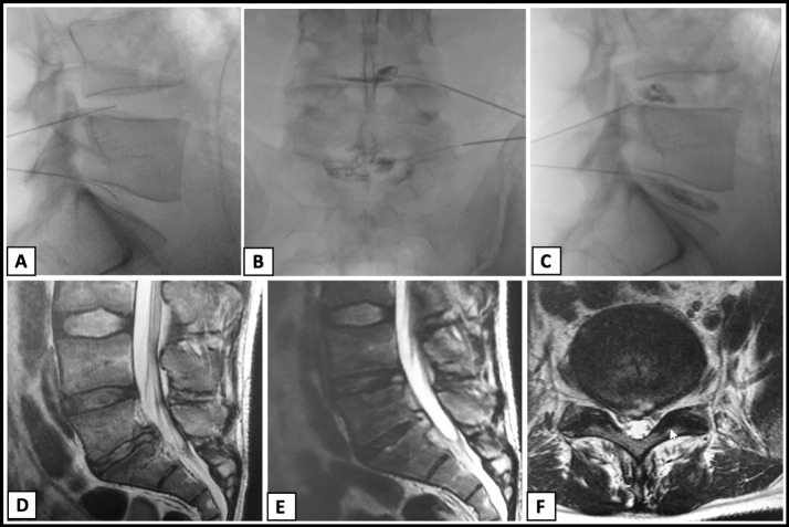
Figure 4.
Case 3. (A) Lateral fluoroscopic image demonstrating L4-L5 and L5-S1 intradiscal needle placement. (B) Anteroposterior fluoroscopic view demonstrating L4-L5 and L5-S needle placement and discogram. (C) Lateral fluoroscopic view demonstrating L4-L5 and L5-S needle placement and discogram. (D) Two weeks post-procedure T2-weighted lumbar magnetic resonance imaging (MRI), sagittal view, demonstrating disc disease with minimal endplate edema at L5-S1. (E) Four weeks postprocedure T2-weighted lumbar MRI, sagittal view, demonstrating increased subchondral edema at the L5-S1 endplates and subtle central bone plate remodeling. (F) Four weeks postprocedure lumbar MRI, axial view, demonstrating large broad-based disc protrusion with associated annular fissure.

