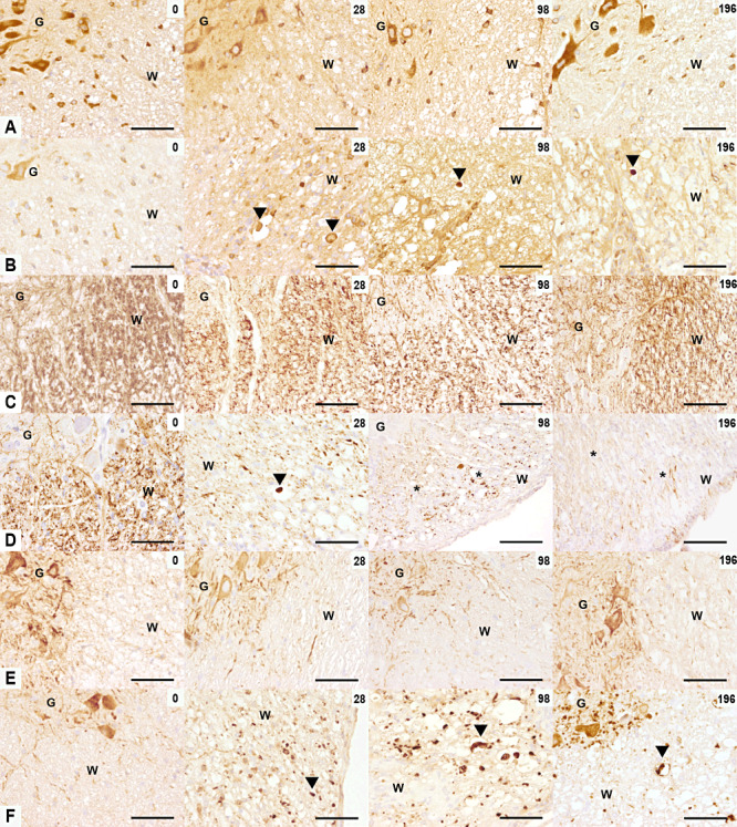
Figure 2.

Amyloid precursor protein (APP), phosphorylated neurofilament (p‐NF), non‐phosphorylated NF (n‐NF) expression in TME. The rows illustrate APP (A,B), p‐NF (C,D), n‐NF (E,F) immunoreactivity in the ventromedial spinal cord of controls (A,C,E) and TMEV‐infected mice (B,D,F) at 0, 28, 98 and 196 dpi. (A,B) At 0 dpi, control and infected mice showed a similar pattern of APP expression in glial cells of the white matter and neurons of the gray matter. From 28 until 196 dpi, APP‐positive spheroids were observed in TMEV‐infected mice (arrowheads). (C,D) At 0 dpi, control and infected mice showed a similar pattern of p‐NF expression with p‐NF‐positive axons in the white matter and p‐NF‐negative neurons in the gray matter. Between 28 and 196 dpi a decreased p‐NF expression was observed in TME. A p‐NF‐positive spheroid is marked with arrowhead and severe axonal loss with asterisks). (E,F) At 0 dpi, control and infected mice showed the same pattern of n‐NF expression with n‐NF‐negative axons in the white matter and n‐NF‐positive neurons in the gray matter. n‐NF axonal expression, marked with an arrowhead, appeared at 28 until 196 dpi in TMEV‐infected mice only. Scale bar = 100 µm. Abbreviations: G = gray matter; TMEV = Theiler's murine encephalomyelitis virus; W = white matter.
