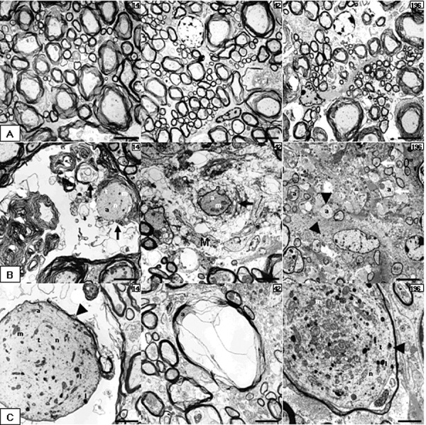
Figure 3.

Electron microscopic features in TME. (A–C) Electron micrographs obtained from spinal cords of placebo (A) and TMEV‐infected mice (B,C) at 14, 42 and 98 dpi. (A) Intact axons at different time points. (B) Specific features of myelin pathology including myelin vesiculation and loss (black arrows, 14 and 42 dpi), naked axons (arrowheads, 196 dpi) and remyelination (white arrows, 196 dpi). (C) Particularities of axonal pathology‐like distended axons (arrowheads) with accumulation of microtubule, neurofilaments, vesiculotubular structures, mitochondria and lysosomes (14 and 196 dpi) or a compressed axon by myelin‐sheath edema (42 dpi). A and B196 scale bar = 4 µm; B14, B42, C14 and C42 scale bar = 2 µm; C196 scale bar = 1 µm. Abbreviations: a = axon; l = lysosomes; m = mitochondria; M = microglia/macrophage cytoplasm; n = neurofilaments; t = microtubules; TMEV = Theiler's murine encephalomyelitis virus.
