
Figure 5.
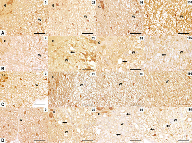
Expression of ubiquitin‐ligated proteins and ubiquitin carboxy‐terminal hydrolase ligase (Uchl‐1) in TME. Ubiquitin (A,B) and Uchl‐1 (C,D) immunoreactivity in the ventromedial spinal cord of control (A,C) and TMEV‐infected mice (B,D) at 0, 28, 98 and 196 dpi. (A,B) At 0 dpi, axonal ubiquitin‐ligated proteins were present in control and infected mice. At 28, 98 and 196 dpi, a lower expression of ubiquitin‐ligated proteins was seen in the TMEV‐infected mice (arrows). (C,D) At 0 dpi, axonal Uchl‐1 was present in the spinal cord of both control and infected mice. At 28, 98 and 196 dpi a lower expression of Uchl‐1 was seen in the TMEV‐infected mice (arrows). Scale bar = 100 µm. Abbreviations: G = gray matter; TMEV = Theiler's murine encephalomyelitis virus; W = white matter.