
Figure 7.
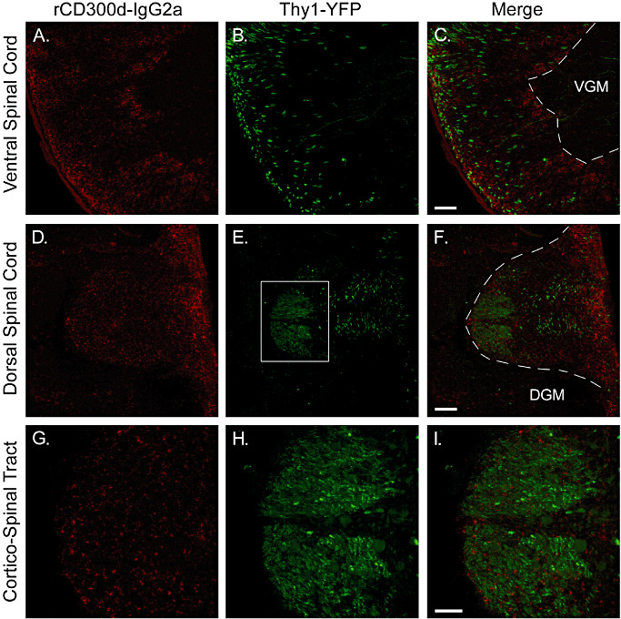
Localization of the putative ligand(s) of rCD300f in spinal cord in vivo. Spinal cord sections from Thy1‐YFP mice were stained with rCD300f‐IgG2a fusion protein, and confocal microscopy was used to visualize specific staining. Corticospinal tract axons can be visualized in green. The red staining for rCD300f‐IgG2a showed a punctate pattern similar to that observed in the brain, and was also mainly present in the white matter tracts of the spinal cord. G–I are higher magnification images from the dorsal corticospinal tract (square area shown in E). No co‐localization was observed between green and red staining. VGM = ventral grey matter; DGM = dorsal grey matter.