
Figure 1.
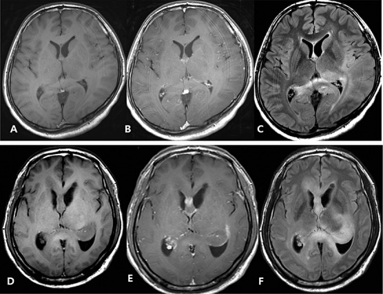
A–C. Diffuse Type 1 gliomatosis cerebri. Pre‐ (A) and postcontrast (B) images show no enhancing portion in the tumor. Flair image (C) indicates diffuse tumor infiltration in left parietotemporal lobe, splenium of corpus callosum, and right parietal lobe with no mass defect and distortion of underlying structures. D–F. Mass forming type 2 gliomatosis cerebri. Pre‐ (D) and Postcontrast (E) shows focal enhancing portion in corpus callosum. Flair image (F) shows tumor infiltrating corpus callosum, left thalamus, basal ganglia and hippocampus with mass formation at hippocampus.