
Fig. 3.
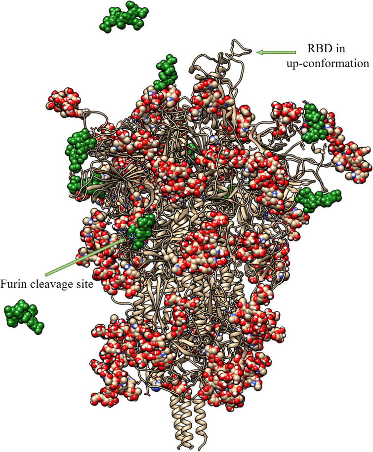
A representative snapshot showing the binding of heparin tetrasaccharides to the glycosylated SARS-CoV-2 S protein, obtained from the unbiased MD simulations. The tan ribbons are the representation of the protein; the glycosylations are the spheres coloured.