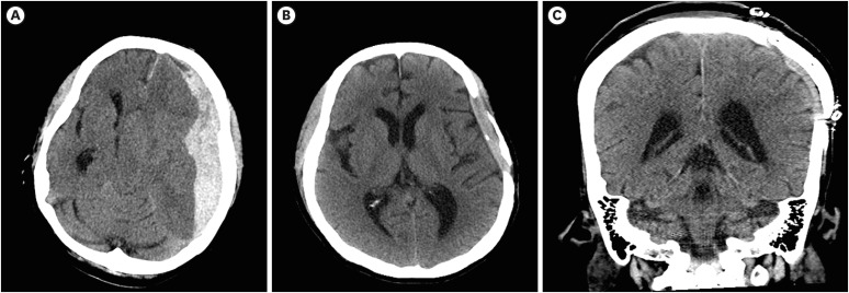
FIGURE 1. (A) Brain CT showing a subdural hematoma in the left fronto-temporo-parietal lobe with midline shifting at the time of injury. Subfalcine and downward transtentorial herniation scans. (B) Absorbed brain hematoma without hydrocephalus 2 years after the accident. (C) Coronal brain CT after cranioplasty.
CT: computed tomography.

