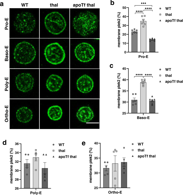
Fig. 4. Early pleckstrin-2 activation and membrane localization during β-thalassemic erythroid precursor differentiation.
a Confocal immunofluorescence microscopy of plek2 (green) localization in sorted (2–3 mice pooled per data point, n = 3 data points per category) WT, β-thalassemic, and apoTf-treated β-thalassemic erythroblasts demonstrate an increased percentage of membrane plek2 localization relative to total plek2 in β-thalassemic relative to: b WT Pro-E and c Baso-E from mice, normalized in apoTf-treated β-thalassemic Pro-E (b) and Baso-E (c) with no differences in: d Poly-E and e Ortho-E. The confocal images were representative optical sections of the cell from at least 50 cells in each condition in 2 different experiments. Scale bar 5 µm. All data are reported as mean ± s.e.m. and p < 0.05 was considered statistically significant (one-way ANOVA) (*p ≤ 0.05, **p ≤ 0.005, ***p ≤ 0.0005). WT = wild type; apoTf = apo-transferrin; plek2 = pleckstrin-2; Pro-E = pro-erythroblasts; Baso-E = basophilic erythroblasts; Poly-E = polychromatophilic erythroblasts; Ortho-E = orthochromatophilic erythroblasts.

