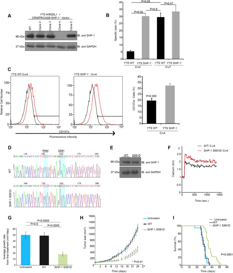
Fig. 1.
Modulation of SHP-1 activity enhances NK cell effector function. a YTS-2DL1 cells were transfected with pSpCas9(BB)-2A-GFP (PX458) and guide RNAs directed against the second exon of SHP-1, and subjected to limiting dilution. Clones were then screened for SHP-1 expression. Clone 4 was selected. b wt YTS-2DL1 or YTS-2DL1 SHP-1−/− cells were incubated with [35S]Met labeled 721-Cw4 or 721-Cw7 target cells at a ratio of 10:1 for 5 h at 37 °C. The specific lysis of target cells was subsequently measured (quantification of different independent experiments is shown, n = 3, mean±SEM). c wt YTS-2DL1 or YTS-2DL1 SHP-1−/− cells were incubated with mCherry expressing 721-Cw4 target cells for 2 h at 37 °C and analyzed for degranulation via FACS (P = 0.005, quantification on the right of independent experiments, n = 4, mean ± SEM). d Sequencing of CRISPR/CAS9 induced knock in of the SHP-1 S591D mutation in YTS-2DL1 cells. Highlighted codons indicate S591 and PAM mutations relative to the wt sample. e YTS-2DL1 wt or YTS SHP-1 S591D cells were lysed and immunoblotted for SHP-1. f YTS-2DL1 wt or SHP-1 S591D cells were loaded with calcium- sensitive Fluo-3-AM, and analyzed for basal intracellular calcium levels for 1 minute. The NK cells were then mixed with 721-Cw4 target cells and incubated at 37°C and analyzed by spectrofluorometry. g, h Tumor growth rate in NOD-Rag1nullIL2rgnull (NRG) mice. NOD-Rag1nullIL2rgnull (NRG) mice were subcutaneously injected with 3 × 10⁶ 721-Cw4 expressing tumor cells. Mice were administered either 3*106 wt YTS-2DL1 or YTS-2DL1 SHP-1 S591D cells by intratumor injections every 3 days. Tumor volumes were calculated as described in the Methods. i Kaplan–Meier curve displaying survival of untreated mice, or mice injected with wt or SHP-1 S591D NK cells. The results represent three independent experiments. For untreated, wt, and SHP-1 S591D injected groups, n = 19, 17, and 19 mice, respectively

