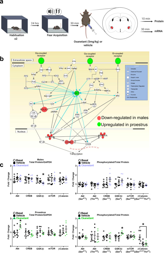
Fig. 5. Osanetant administered 30 min after FA reduces Akt/GSK3β/β-Catenin pathway activation in males and it enhances it in proestrous females in the amygdala.
a Fear procedure to obtain amygdala tissue for biochemical studies. b Schematic representation of the involved PI3K/Akt pathway revealed by the G-Coupled Protein Receptor (GCPR) qPCR array results after bioinformatics analysis of the Ingenuity Pathway Analysis (IPA) software. Red indicates downregulation and green upregulation (n = 4 per group). c Western blot analyses of the Akt/GSK3β/β-Catenin signaling pathway in amygdalar lysates of males in males (basal: n = 7; vehicle: n = 8; osanetant: n = 8) revealed downregulated total GSK3β expression in osanetant treated mice compared to vehicle (p = 0.006) and basal (p = 0.011) groups; while proestrous females (basal: n = 6; vehicle: n = 5; osanetant: n = 5) showed increased total Akt expression in osanetant treated mice compared to vehicle (p = 0.035) and basal groups (p = 0.029), with a decrease in p-β-Catenin in osanetant treated females compared to basal animals (p = 0.017). Original blots and scans are shown in Supplementary Fig. 6. Protein levels were normalized to GADPH (left graphs) and phosphorylated proteins were normalized to total protein levels (right graphs). Data are mean ± SEM. *p ≤ 0.05 vs. the other groups. For qPCR analysis, two-tailed t-test or Mann–Whitney’s U test were used for statistical analyses. Western blots were analyzed with one-way ANOVA or Kruskal–Wallis’ χ2 test. Pairwise comparisons are indicated when appropriate.

