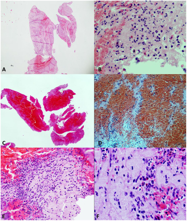
Figure 3.

Different samples of clots. Hematoxylin–eosin (HE) stain. (A) (HE2×) and (B) (HE20×). Platelet fibrin-predominant clot (FPC) with moderate number of complete neutrophils. (C) (HE2×) and (D) (HE20×). Red cell-predominant clot (RPC), with poorly formed Zahn lines. (E) (HE20×) and (F) (HE40×). Mixed clot (MC) with plenty of neutrophils, most of them of degenerated appearance.
