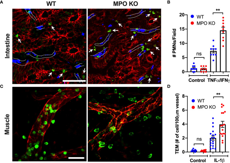
Figure 1.
PMN tissue localization is enhanced in the absence of functional MPO. (A, B) Inflammation of the intestinal mucosa was induced by intraperitoneal administration of TNFα and IFNγ (500ng, each 24hr). Immunofluorescence analyses were performed on OCT-frozen 10µm-sections. PMNs, intestinal epithelial cells and blood vessel were visualized by S100A8 (green), E-Cadherin (red) and PECAM (CD31, blue) staining respectively. (A) Representative images and (B) quantification show increased number of tissue-infiltrating PMNs in MPO KO mice. Blood vessel locations are highlighted by dotted lines. White arrow point to tissue localized PMNs. (C, D) Inflammation of the cremaster muscle was induced by intrascrotal administration of IL-1β (50ng, 4hr). PMNs and ECs were visualized in a whole mount muscle preparation by respective staining with S100A8 (green) and CD31 (red). Consistently, PMN numbers in tissue were significantly elevated in MPO KO animals. N=3 independent experiments with 3 mice per condition. **p < 0.01. ns, not significant.

