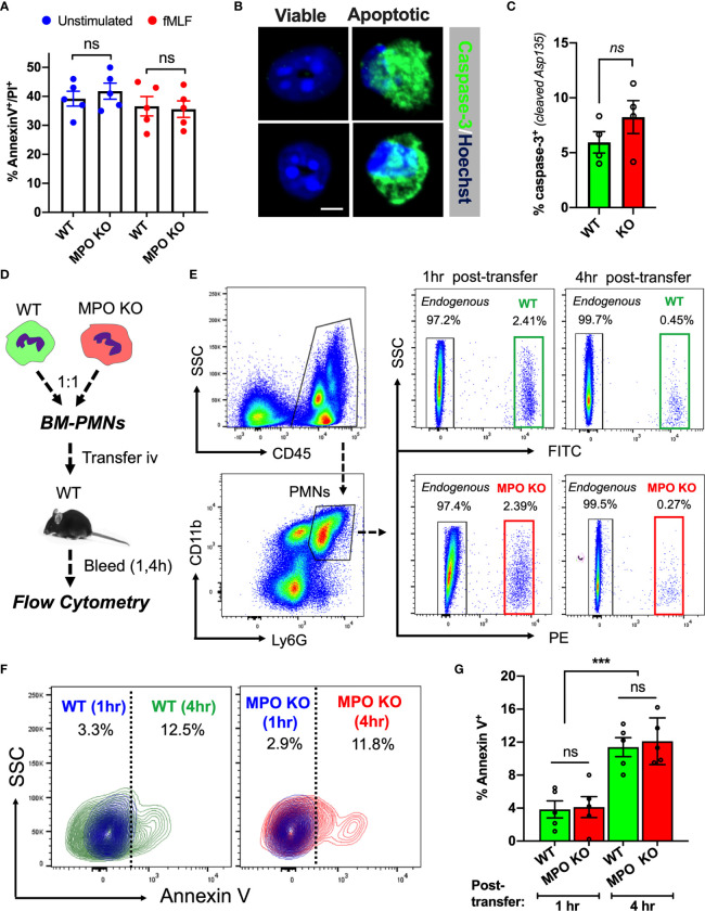
Figure 2.
MPO expression does not impact PMN viability. (A) PMN viability was assessed in ex vivo cultured BM-derived PMNs with/without fMLF-stimulation using Annexin V/PI staining and flow cytometry. Data shown as percent Annexin V/PI positive cells at 24hr in cultures. No significant differences between MPO KO and WT mice were observed. N=5 independent experiments. (B, C) Representative images and quantification of cleaved caspase-3 in fMLF-stimulated BM-derived WT and MPO-KO PMNs. No significant differences between MPO KO and WT mice were observed. N=4 independent experiments. The bar is 5µm. (D–G) PMN viability was examined in vivo in adoptively transferred PMNs. (D) Schematic depicting experimental timeline. Freshly isolated murine WT and MPO KO BM-PMNs were respectively labeled with green and red fluorescence (CellTracker) and injected intravenously at a 1:1 ratio into WT recipients that were pre-stimulated with IL-1β (50ng, 1hr, ip., to induce systemic inflammation). Annexin V staining and flow cytometry was used to gauge viability of transferred and endogenous PMNs at 1 and 4hr. (E–G) Representative flow diagrams of the gating strategy to identify transferred and apoptotic WT and MPO KO PMNs and quantification of apoptotic (AnnexinV-positive) PMNs in the circulation. No significant difference in viability between MPO KO and WT PMNs. N=5 mice per condition. ns, not significant. ***p < 0.001.

