Figure 5.
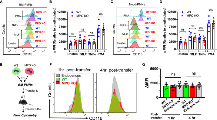
CD11b surface expression is not altered by the genetic deletion of MPO. (A–D) CD11b expression was assessed by flow cytometry in BM-derived and circulating blood PMNs with/without ex vivo inflammatory stimulation. (A, B) Representative flow diagram and quantification of CD11b expression in BM-PMNs. (C, D) Representative flow diagram and quantification of CD11b expression in circulating PMNs. N=5 independent repeats per condition. (E–G) CD11b expression was assessed in fluorescently labeled WT (green) and MPO KO (red) PMNs at 1 and 4hr following adoptive intravenous transfer into IL-1β-stimulated (50ng, 1hr) WT recipient mice. (E) Schematic depicting experimental time line. (F, G) Representative flow diagrams and quantification reveal no significant differences in CD11b expression. N=5 mice per condition. ns not significant.

