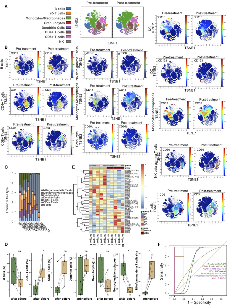
Figure 1.
The immune landscape of the kidney. (A) t-SNE plots of CD4+ T cells, CD8+ T cells, B cells, NK cells/γδ T cells, monocytes/macrophages, granulocytes, and dendritic cells. (B) t-SNE plots of CD19, CD3, CD4, CD8a, CD56, gdTCR, CD33, CD14, CD66b, CD11c, and CD123 expression in the pre- and posttreatment groups. (C) The proportions of main cell types in all samples. (D) Comparison of the proportions of main cell types between the pre- and posttreatment groups. (E) Heatmap of the marker expression for all samples. (F) ROC curve of the main cell types predicting the pre- and posttreatment groups. *P<0.05; ns, not significant.

