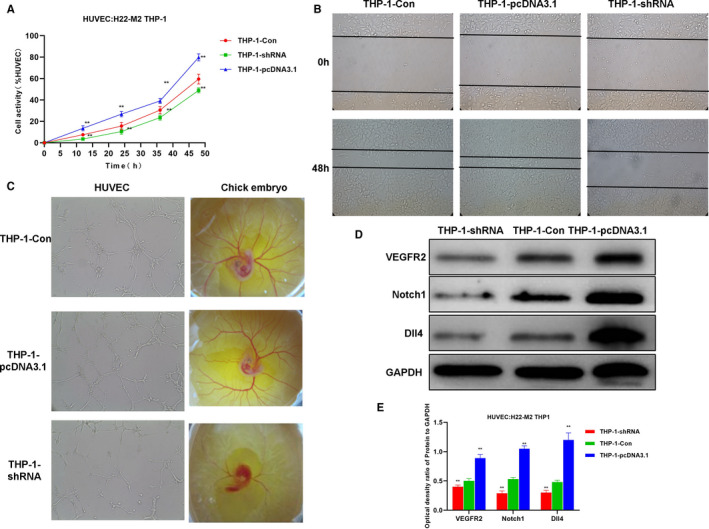
FIGURE 6.

H22‐induced M2 macrophages‐induced angiogenesis. A, HUVEC cell viability results (n = 5): The cell viability of the THP‐1‐pcDNA3.1 group was significantly higher than that of the THP‐1‐Con. Comparison with THP‐1‐Con, *P <.05; **P <.01. B, HUVEC cell migration assay (n = 5): The cell migration capacity of THP‐1‐pcDNA3.1 group was significantly enhanced than that of THP‐1‐Con group. C, In vitro tube formation assay of HUVEC and chick embryo chorioallantoic membrane assay (n = 5): The vessel formation ability of THP‐1‐pcDNA3.1 group was significantly enhanced than THP‐1‐Con group. D and E, Expression of key signal proteins for angiogenesis (n = 3): The protein expression of Notch1, Dll4 and VEGFR2 in the THP‐1‐pcDNA3.1 group was significantly higher than that of the THP‐1‐Con group, and the expression in the HP‐1‐shRNA group was lower than that of the THP‐1‐Con group. Comparison with THP‐1‐Con, *P <.05; **P <.01
