FIGURE 2.

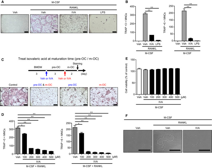
IVA blocks OC differentiation induced by RANKL. A, B, BMDMs were stimulated with IVA (200 μM) or LPS (1 μg/mL) under OC differentiation conditions (30 ng/mL M‐CSF plus 100 ng/mL RANKL) for 5 days. TRAP+ MNCs (>3 nuclei or >5 nuclei) were counted and considered to be OCs. C, A protocol to test the effects of IVA on OC generation at different time points (top). IVA was added at day 3 of BMDMs and/or at day 6 of pre‐OCs during osteoclastogenesis. Mature OCs were stained by TRAP staining solution (bottom). D, BMDMs were stimulated with IVA (0, 100, 200, 300, 400, and 500 μM) under OC differentiation conditions (30 ng/mL M‐CSF plus 100 ng/mL RANKL) for 5 days. With TRAP staining, the TRAP+ MNCs (>3 nuclei or >5 nuclei) were counted and considered to be OCs. (E) BMDMs were stimulated with IVA (0, 100, 200, 300, 400, and 500 μM) in the presence of M‐CSF (30 ng/mL) for 3 days. After harvesting the supernatant, the levels of LDH released were monitored by measuring ODs at 490 nm. (F) Representative images of bone resorption area on the Corning Osteo Assay Plate. BMDMs were stimulated with M‐CSF (30 ng/mL) and RANKL (100 ng/mL) with or without IVA (200 μM) for 7 days. Data are representative of three independent experiments (A, C, F). Data are presented as the mean ± SEM (n = 3 for B, D, E). * P < .05, ** P < .01. Scale bar, 2 μm (A, C bottom). Scale bar, 100 μm (F)
