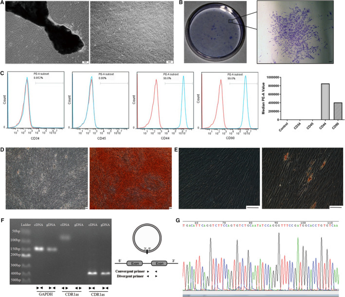
FIGURE 1.

Identification and validation of PDLSCs and CDR1as. A. PDLSCs were derived from PDL explants on day 7 and cultured in normal medium until passage number 3. B. Single colonies formed after PDLSCs were cultured for 10 days. C. PDLSCs were characterized by detection of mesenchymal stem cell surface markers (CD44 and CD90) through flow cytometric analysis. Leukocyte markers (CD34 and CD45) were used as a negative control. D. Alizarin Red staining showing the mineralized matrix of osteo‐induced PDLSCs. E. Oil Red O staining showing the oil deposition in adipo‐induced PDLSCs F. Divergent and convergent primers of CDR1as were designed to specifically target the circular junction site and the linear region of CDR1as, respectively, for qRT‐PCR. The circular structure of CDR1as was validated by agarose gel electrophoresis. G. The head‐to‐tail splicing of the CDR1as RT‐PCR product was confirmed by Sanger sequencing. Scale bar (A, B, D, E), 100 μm
