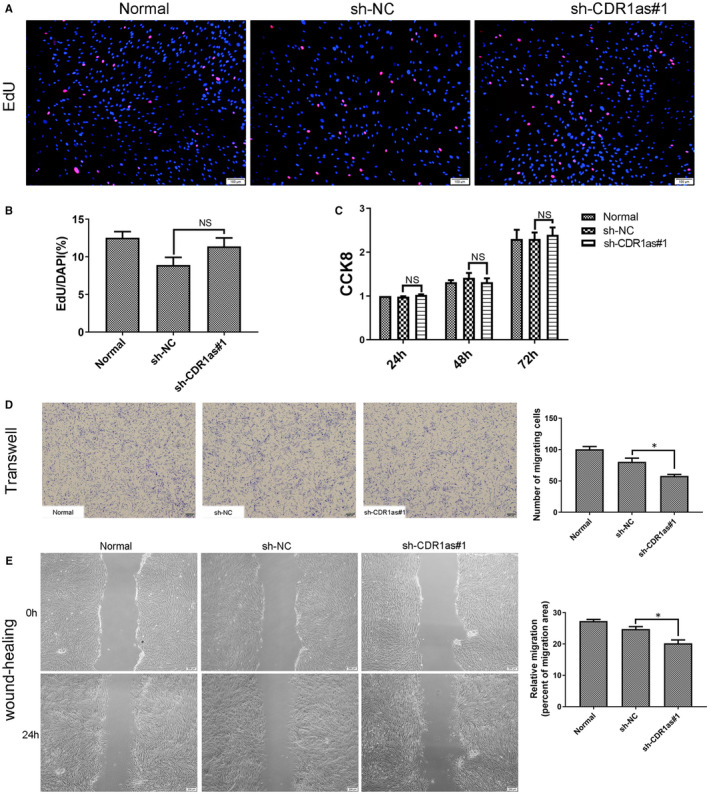
FIGURE 3.

Knockdown of CDR1as inhibits migration and wound healing capacities of PDLSCs. A. DNA synthesis of PDLSCs was assessed by EdU assay after transfection with sh‐CDR1as#1 and sh‐NC for 48 h. B. Quantitative EdU assay data from A. C. Proliferation of PDLSCs in three groups was assessed at 24 h, 48 h and 72 h using a CCK8 kit. D. Migration ability of PDLSCs in three groups was assessed by transwell assay. Cells that migrated to the underside of the membrane were stained and counted. E. The average wound widths at 0 h and 24 h were analysed to assess the wound healing capacity of PDLSCs. Scale bar (A), 100 μm. Scale bar (D, E), 200 μm. Quantitative data are presented as mean ± SD. *P < .05; NS, not significant, by Student's t test
