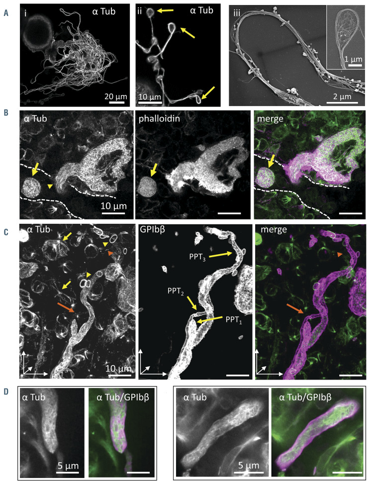
Figure 4.
Microtubules are differentially distributed along proplatelets in situ compared to in vitro. (A) i-ii: cultured proplatelets (cPPT) from wild-type (WT) megakaryocytes (MK) differentiated in culture for 4 days, labeled with an antibody against α tubulin. iii: microtubule bundles within the cPPT shaft and coils in the bud (inset), observed by scanning electron microscopy (SEM) after the removal of the membrane by treatment with Triton X-100. (B) Confocal images of a nascent native proplatelets (nPPT) within bone marrow 30-mm thick sections revealing an absence of microtubule bundle organization (antibodies against α tubulin in green) and F-actin (phalloidin labeling, in magenta). Dotted lines represent sinusoid vessel. Note the presence of an nPPT transversally sectioned (arrow) in the sinusoid, again lacking microtubule bundles. (C-E) In situ bone marrow nPPT immunolabeling with antibodies against α tubulin (green) and GPIbβ (magenta). (C) 3D visualization of z-stack (30 mm thick) showing parts of three entangled nPPT after GPIbb labeling, denoted PPT1, PPT2 and PPT3. Tubulin labeling was discontinuous being weak to absent in certain portions of the nPPT. For example, labeling becomes weaker in the upper part of the longest nPPT (PPT3) (orange arrowhead). Tubulin labeling in the thinnest nPPT (PPT2) was hardly visible in our settings (orange arrow). Marginal band of platelets (yellow arrowheads) and mitotic spindles (yellow arrows) were well labeled denoting well-preserved microtubules. (D-E) Confocal single-plane images at higher magnification showing nPPT portions. Note that the microtubules are not arranged longitudinally. (B-E) Representative of four wild-type (WT) femur bone marrow from two mice.

