Figure 2.
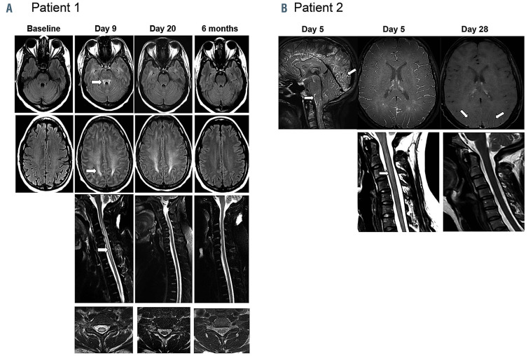
Leucoencephalomyelopathy changes on magnetic resonance imaging in patient 1 and 2. (A) Patient 1: T2-weighted fluid attenuation inversion recovery axial images of the brain (top 2 rows) demonstrate evolution and resolution of symmetrical T2 hyperintensity within the superior cerebellar peduncle, the centrum semiovale white matter with respect of U-fibers, from day 9 to 6 months. T2 fast spin echo (FSE) with fat saturation sagittal imaging of the cervico-thoracic spine (row 3) and representative T2 FSE axial sections of the cervical cord (row 4) illustrate resolution of extensive centromedullary T2 changes over time. By 6 months, magnetic resonance imaging (MRI) changes normalized. (B) Patient 2: brain and cervical spine MRI performed at neurotoxicity onset on day 5 demonstrating diffuse infratentorial and supratentorial leptomeningeal uptake predominant around the brain stem, cervical spinal cord, and cerebellar and occipital sulci (3D FLAIR with gadolinium). Diffuse leptomeningeal enhancement (3D T1 Black Blood with gadolinium) and T2 hyperintensity extending from C2 to C4 was also noted with a slender swelling of the spinal cord. At day 28 re-evaluation, MRI showed almost complete resolution of leptomeningeal uptake with persistence of discreet gadolinium enhancement in the posterior parieto-occipital sulcus (3D T1 VISTA with gadolinium) and complete disappearance of the intramedullary T2 hyperintensity.

