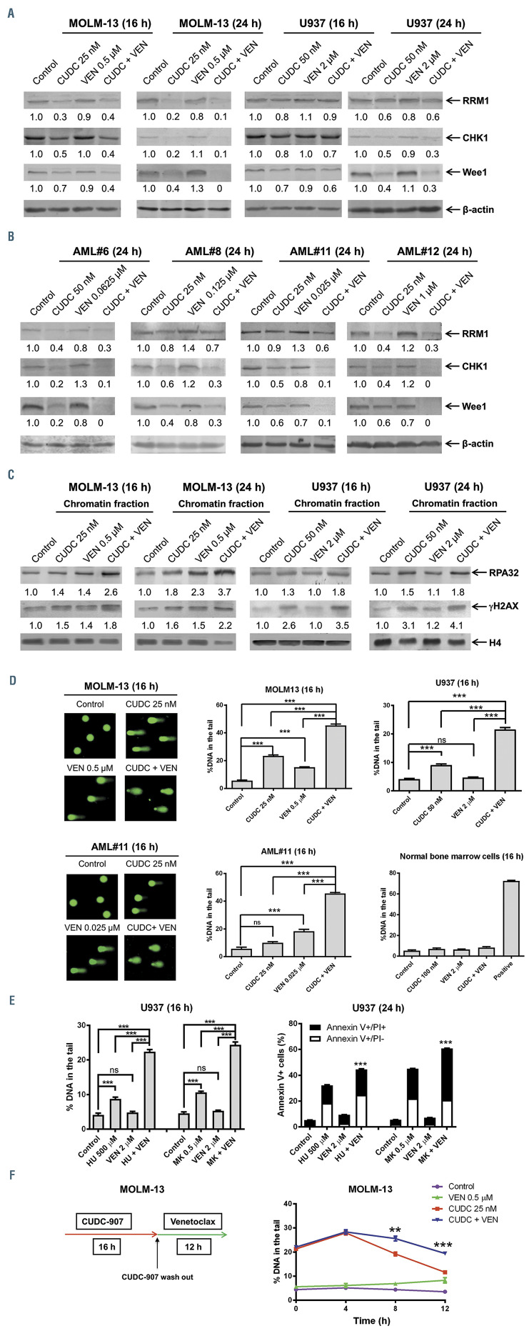
Figure 4.
Venetoclax enhances CUDC-907-induced DNA damage in acute myeloid leukemia cells. (A, B) Acute myeloid leukemia (AML) cells were treated with vehicle control, venetoclax, CUDC-907, or in combination for 16 or 24 hours, and whole cell lysates were subjected to western blotting. Representative western blots are shown. The fold changes for the densitometry measurements, normalized to b-actin and then compared to no drug control, are indicated below the corresponding blot. (C) AML cells were treated as in panels A, B. The levels of RPA32 and γH2AX bound to chromatin were analyzed by western blotting. Densitometry measurements normalized to histone H4 and then compared to vehicle control are presented below the corresponding blot. (D) AML cells and normal bone marrow cells were treated for 16 hours with vehicle control, venetoclax, CUDC-907, venetoclax + CUDC-907, or a positive control (20 mM daunorubicin for 4 hours). Representative alkaline comet assay images for MOLM-13 and AML#11 are shown. Representative images for U937 and normal bone marrow cells are shown in the Online Supplementary Figure S4. Data are graphed as median percent DNA in the tail from three replicate gels ± standard error of the mean (SEM). ns indicates not significant and ***P<0.001. (E) U937 cells were treated with venetoclax in the presence or absence of hydroxyurea (HU) or MK-1775 (MK) for 16 hours and then subjected to the alkaline comet assay (left panel). Data are graphed as median percent DNA in the tail from three replicate gels ± SEM. ns indicates not significant and ***P<0.001. Representative images are shown in the Online Supplementary Figure S5. The treated U937 cells were also subjected to Annexin V/PI staining and flow cytometry analysis (right panel). Mean percent Annexin V+ cells ± SEM are shown. ***P<0.001 compared to single drug treatments. (F) MOLM- 13 cells were treated with or without CUDC-907 for 16 hours. The cells were washed with PBS three times and then split, half receiving fresh media and the other half receiving fresh media plus venetoclax. Cells were collected at 0, 4, 8, and 12 hours after addition of venetoclax. Alkaline comet assay results are shown as median percent DNA in tail from three replicate gels ± SEM. **P<0.01 and ***P<0.001 compared to CUDC-907 treatment. Representative images are shown in the Online Supplementary Figure S7.

