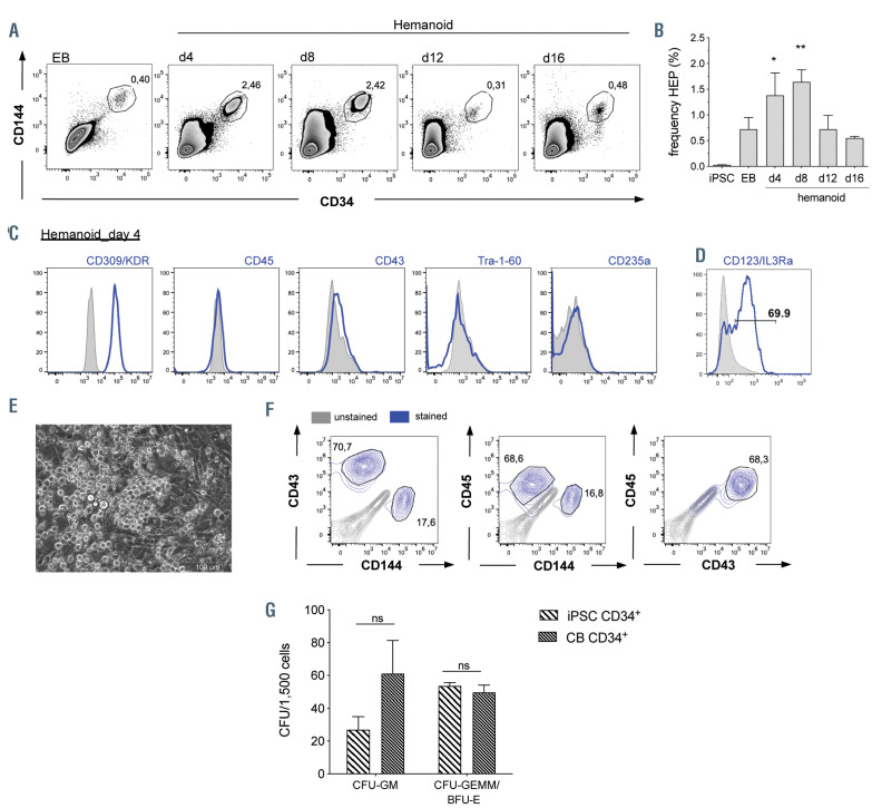
Figure 3.
Characterization of hemato-endothelial progenitors within hemanoids. (A and B) Frequency of CD34+/CD144+ hemato-endothelial progenitors (HEP) at different days of hematopoietic specification. (A) Representative flow cytometry data (percentage of gated population is indicated) and (B) quantification of n=4, mean±standard error of the mean (SEM). (C) Detailed surface marker phenotype of HEP derived from hemanoids day 4 analyzed by flow cytometry (pre-gated on CD34+/CD144+ cells, histogram overlay: unstained grey filled, surface marker expression blue, representative data from n=3, approx. 1,000 HEP were analyzed per sample). (D) Expression of the IL3Ra/CD123 on CD34+/CD144+ HEP (histogram overlay: unstained grey filled, surface marker expression blue, representative data from n=3, approx. 1,000 HEP were analyzed per sample, percentage of gated population is indicated). (E) Morphology of HEP after 1 week in endothelial to hematopoietic transition (EHT) culture (scale bars: 100 mm and 200 mm, respectively). (F) Flow cytometry analysis of CD144, CD43 and CD45 expression on CD34+ cells after 1 week EHT culture (unstained: grey, surfacemarker expression: blue, representative data from n=2, approx. 10,000 CD34+ cells were analyzed per sample, percentage of gated population is indicated, pre-gating on CD34 is shown in the Online Supplementary Figure S3C). (G) Left: Frequency of colony forming units (CFU) of iPSC-derived CD34+ cells after 1 week of EHT culture and cord blood-derived CD34+ cells (n=4, two biological and two technical replicates, mean±SEM). *P<0.05, **P<0.01, ns: not significant; statistical significance was assessed using (B) One-way ANOVA with Dunnetts multiple comparison test and (G) two-way ANOVA with Sidak's multiple comparisons test.)

