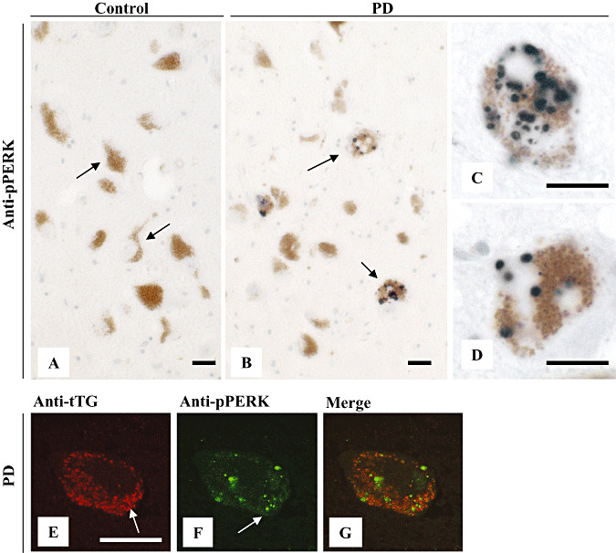
Figure 5.

Phosphorylated pancreatic ER kinase (pPERK) staining does not colocalize with tissue transglutaminase (tTG)‐immunoreactive granules in melanized neurons in Parkinson's disease (PD) brains. Immunohistochemical staining of pPERK (antibody Sc‐32577) was absent in melanized neurons of the substantia nigra (SN) in control brain (A). In contrast, pPERK staining was observed in melanized neurons of the SN in PD brains (B–D). Double immunofluorescence of tTG (antibody 06‐471) (E, arrow) and pPERK staining (antibody Sc‐32577) (F, arrow) demonstrated no colocalization of pPERK in tTG‐immunoreactive granules in melanized neurons (G). Scale bars: (A–G) 20 µm.
