
Figure 3.
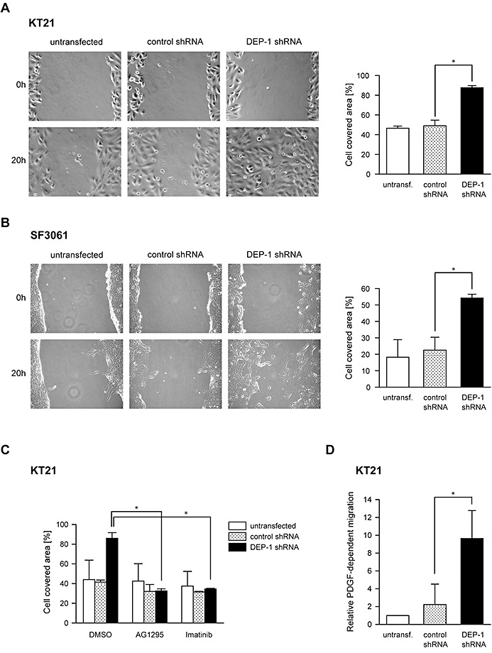
DEP‐1 is a negative regulator of meningioma cell migration. A. Untransfected KT21 and SF3061 cells and B. Cells stably expressing DEP‐1‐targeting shRNA or control shRNA were subjected to wounding assays. Images of three positions were taken 0 h and 20 h after wounding, and the area of the wound covered with cells after 20 h was quantified (mean values ± SD of three independent experiments, *P < 0.05 by t‐test). C. Untransfected, DEP‐1‐targeting shRNA or control shRNA expressing KT21 cells were treated as in A, except that the PDGF receptor kinase inhibitors AG1295 (20 µM) or Imatinib/STI571 (2 µM), or the solvent DMSO (as indicated) were added to the medium after wounding the cell layers (mean values ± SD of two independent experiments, *P < 0.05 by t‐test). D. Untransfected, DEP‐1‐targeting shRNA or control shRNA expressing KT21 cells were serum‐starved and subjected to a chemotaxis assay using a modified Boyden chamber. Cells that migrated towards a PDGF‐BB gradient to the lower side of the membrane were counted (mean values ± SD of three independent experiments, *P < 0.05 by t‐test).