
Figure 5.
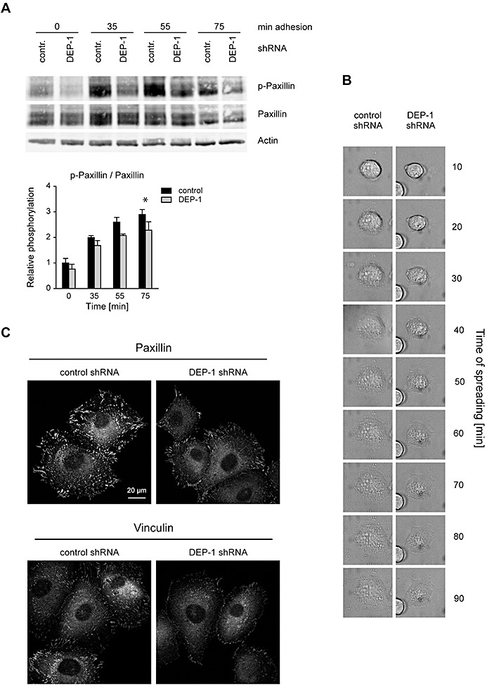
DEP‐1 positively regulates paxillin phosphorylation, and enhances cell spreading and formation of focal adhesion complexes. A. KT21 cells stably transfected with DEP‐1‐targeting or control shRNA expression constructs were starved in suspension and seeded in fibronectin‐coated plates. After the indicated times, adherent cells were lysed, and lysates were analyzed by immunoblotting. Equal loading was verified by detection of β‐actin. The blots were quantified by densitometry, and intensities of p‐paxillin normalized to paxillin signals are displayed in the graph (mean values ± SEM of four independent experiments). Two‐way ANOVA and Bonferroni's multiple comparison test was applied to test for significant differences. Signaling intensities of DEP‐1 shRNA vs. control shRNA expressing cells differed significantly with respect to the whole time‐series of stimulation (**P < 0.01). Comparisons for individual time points displayed in the graphs were also statistically tested (*P < 0.05) B. Reduced spreading of DEP‐1‐depleted KT21 cells. Attachment of the indicated cell lines was examined simultaneously under a time‐lapse microscope over a period of 90 min. A representative example is shown. C. KT21 cells stably transfected with DEP‐1‐targeting or control shRNA expression constructs were starved in suspension in serum‐free medium for 1 h and seeded on fibronectin‐coated cover slips. After 2 h, cells were fixed and stained with paxillin or vinculin antibodies followed by Cy3‐conjugated secondary antibodies. Images were taken using a laser scanning microscope and converted into grayscale. A representative example is shown.