
Figure 7.
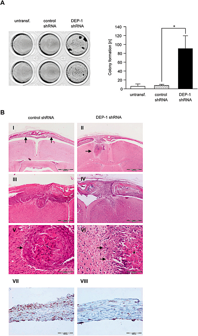
DEP‐1 knockdown promotes meningeal cell transformation and invasion. A. Parental KT21 cells, KT21 cells stably transfected with non‐targeting shRNA, or with DEP‐1‐targeting shRNA, as indicated, were grown in soft agar for 15 days. Colonies were stained with MTT and counted. An example and quantification of three independent experiments with quadruplicate determination are depicted. B. KT21 cells stably transfected with non‐targeting shRNA, or with DEP‐1‐targeting shRNA as indicated were stereotactically transplanted into the prefrontal cortex of NMRI nu/nu mice. Tumor growth and brain invasion were analyzed histologically. Scale bars represent 1000 µm (I, II) 500 µm (III, IV), and 100 µm (V–VIII). V and VI are higher magnifications of III and IV, respectively. The arrows indicate tumor tissue in (I) and brain invasion of tumor cells in (II). While KT21 meningioma cells bearing control shRNA show a more displacing mode of brain invasion (III; arrow in V), DEP1‐depleted KT21 cells were characterized by a more diffuse type of brain invasion (IV; arrows in VI; asterisk marks tumor necrosis). Immunostaining of paraffin‐embedded intracranial tumors derived from control (VII) or DEP‐1 knockdown (VIII) cells using an anti‐DEP‐1 antibody revealed maintenance of the DEP‐1 status throughout the experiment (scale bar 100 µm).