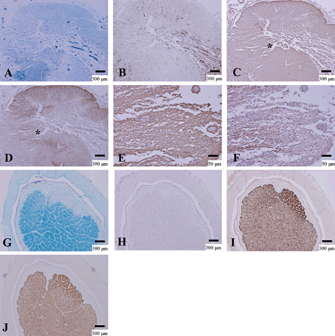
Figure 2.

Upregulation of AQP4 in chronic NMO lesions. (A–F) Serial sections of chronic active demyelinating lesions in the spinal cord of NMO‐10 representing Pattern C and N. A. The spinal cord has irregularly‐shaped demyelinating lesions with necrosis and cavity formation. Reactive proliferation of capillary vessels is noted. B. CD68‐positive macrophages are still abundant in the perivascular regions. C. The lesion is immunopositive for GFAP except for in the cavity center. D. Upregulation of AQP4 in extensively demyelinated lesions. E. High magnification in the lesion indicated by the asterisk in C shows increased GFAP immunoreactivity. F. The same area as E demonstrates AQP4 staining in areas of astrogliosis. (G–J) Serial sections of chronic inactive demyelinating lesions in the optic nerve from NMOSD, representing Pattern D. G. Sharply demarcated demyelinating plaque in the optic nerve. H. Macrophage infiltration is absent. I. GFAP‐positive chronic astrogliosis covers the demyelinating plaque. J. AQP4 expression is upregulated in the areas of chronic astrogliosis. A, G, KB staining; B, H, CD68 immunohistochemistry (IHC); C, E, I, GFAP IHC; D, F, J, AQP4 IHC. Scale bar = 300 µm (A–D, G–J); 50 µm (E, F). AQP4 = aquaporin‐4; GFAP = glial fibrillary acidic protein; KB = Klüver‐Barrera staining; NMO = neuromyelitis optica; NMOSD = NMO spectrum disorder.
