
Figure 3.
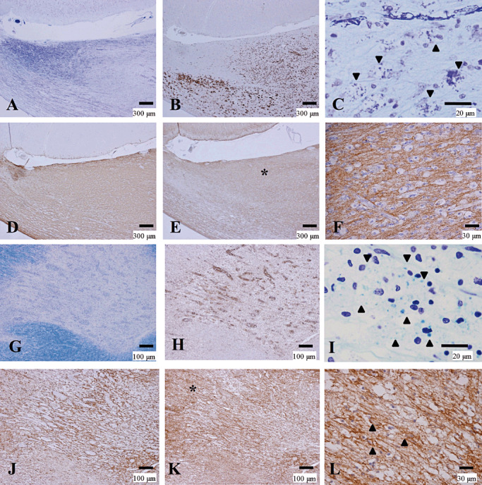
Preserved AQP4 immunoreactivity in actively demyelinating NMO lesions. (A–F) Serial sections of an actively demyelinating lesion from NMO‐6 representing Pattern D. A. Demyelinating plaque in the corpus callosum. B. The lesion is infiltrated by numerous foamy macrophages stained with CD68. C. Macrophages containing myelin debris (arrowheads). D. The lesion shows strong GFAP immunoreactivity. E. The same area as in D, with AQP4 expression as strong and diffuse as that of GFAP. F. High magnification of the area indicated by the asterisk in E. AQP4 expression is preserved despite dense macrophage infiltration. (G–L) Serial sections of an optic nerve lesion from NMO‐4 representing Pattern D. G. Sharply demarcated demyelinating plaques are seen. H. The optic chiasm is densely infiltrated with foamy macrophages. I. Numerous macrophages contain myelin debris. J. Numerous GFAP‐positive reactive astrocytes in the lesion center and the surrounding areas. K. AQP4 immunoreactivity is enhanced at the lesion center. L. High magnification of the area indicated by the asterisk in K demonstrates AQP4 staining along the entire processes and outlining the cytoplasm of hypertrophic reactive astrocytes (arrowheads). A, C, G, I, KB staining; B, H, CD68 immunohistochemistry (IHC); D, J, GFAP IHC; E, F, K, L, AQP4 IHC. Scale bar = 300 µm (A, B, D, E); 100 µm (G, H, J, K);30 µm (F, L); 20 µm (C, I). AQP4 = aquaporin‐4; GFAP = glial fibrillary acidic protein; KB = Klüver‐Barrera staining; NMO = neuromyelitis optica.