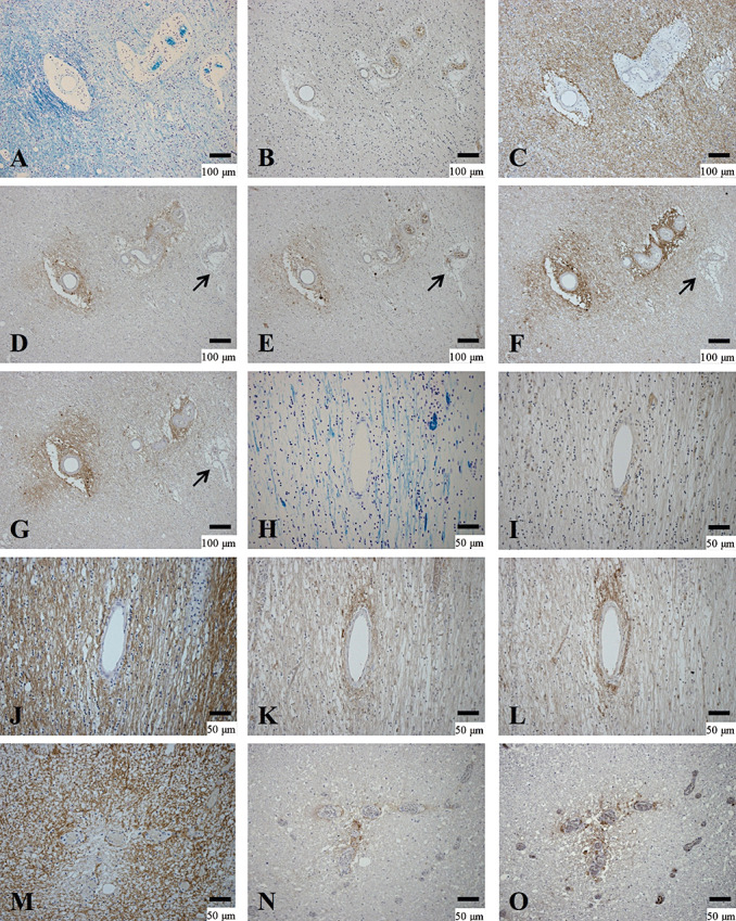
Figure 6.

Heterogeneity between perivascular deposition of activated complement and immunoglobulins and AQP4 expression. (A–G) Serial sections of the peripheral area of chronic active lesions in the cerebral white matter of NMO‐10. A. Demyelinating lesions confined to the vicinity of blood vessels. B. No inflammatory cell infiltration. C. AQP4 immunoreactivity on astrocyte processes. Immunoreactivities to activated complement (C3d (D) and C9neo (E)) and immunoglobulins (IgM (F) and IgG (G)) in the perivascular areas. Note that one blood vessel does not show deposition of activated complement or immunoglobulins (arrow in D–G). (H–L) Serial sections in the cerebral white matter of the same case. H. A chronic inactive demyelinating lesion. I. Few CD68‐positive macrophages in the lesion. J. AQP4 labeling of the astrogliosis covering the lesions. C3d (K) and IgG (L) immunoreactivities are most intense in the perivascular area. (M–O) Serial sections of a chronic active demyelinating lesion in the spinal cord of the same case. M. Loss of AQP4 immunoreactivity is confined to the perivascular areas associated with C3d (N) and IgM (O) deposition. A, H, KB; B, I, CD68 immunohistochemistry (IHC); C, J, M, AQP4 IHC; D, K, N, C3d IHC; E, C9neo IHC; F, O, IgM IHC; G, L, IgG IHC. Scale bar = 100 µm (A–G); 50 µm (H–O); 15 µm (C). AQP4 = aquaporin‐4; GFAP = glial fibrillary acidic protein; KB = Klüver‐Barrera staining; NMO = neuromyelitis optica.
