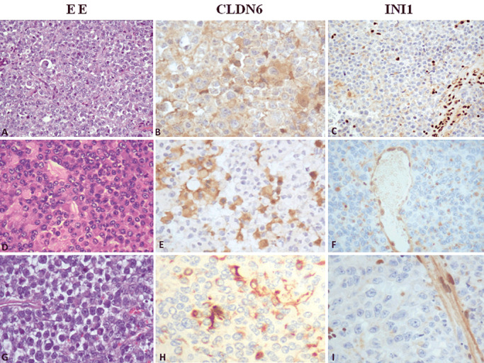
Figure 1.

Histopathology and evaluation of claudin‐6 and SMARCB1 (INI1) immunohistochemistry in atypical teratoid rhabdoid tumor (AT/RT). A–C. Medium‐power view illustrates a typical AT/RT, with loss of nuclear staining in tumor cells. Neoplastic cells show and strong staining for claudin‐6 (score 3+). D–F. A case of AT/RT, negative for INI1 protein and with a moderate staining for claudin‐6 (score 2+), original magnification 200×. G–I. A case of AT/RT with absence of INI1 protein and with rare cells positive for claudin‐6, original magnification 200×.
