
Figure 2.
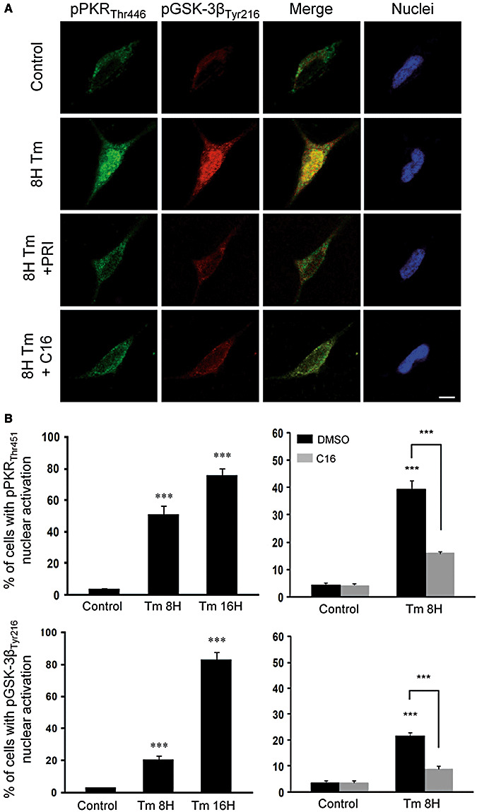
Effect of PKR inhibitors (PRI and C16) on tunicamycin (Tm) induced activation of PKR and GSK‐3β in SH‐SY5Y cells. A. Untreated neuroblastoma cell lines showing slight staining of pPKRThr446 (green) and pGSK‐3βTyr216 (red) in the cytoplasm, without apoptotic nuclei. After 8 h of Tm (5 µg/mL) treatment, the labeling of pPKRThr451 and pGSK‐3βTyr216 and their co‐localization were increased in the cytoplasm and nuclei. Adding PRI (50 µM) or C16 (1 µM) after 8 h of Tm exposure lead to an attenuation of the activation of PKR and GSK‐3β in the cytoplasm and nucleus associated with a strong reduction of co‐localization. Horizontal bar 10 µM. B. The cell counting confirmed that PKR (50%) and GSK‐3β (20%) activated after 8 h of Tm treatment and it increases, respectively, to 75% and 80% after 16 h. C16 attenuates both nuclear activation of PKR and GSK‐3β.