
Figure 3.
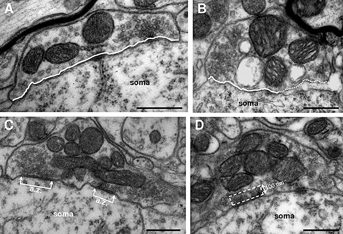
Ultrastructural characterization of hypoglossal motoneurons‐attached boutons. A, B. Two boutons with flat/pleomorphic (F/P) vesicles attached to the motoneuron soma from wild‐type (WT) non‐transgenic littermates (A) and hSOD1G93A (B) mice. Note the enlarged subsynaptic space observed in the bouton from the transgenic mouse. Continuous and dotted lines indicate the segment of bouton attached and detached, respectively, from the motoneuron cell body. C. A F/P‐type bouton with two active zones (a.z.) is shown. The distance between connected arrows represents the length of each a.z. Note that an end of the bouton is detached from the motoneuron. D. Picture illustrating the rectangular area considered in counting the number of vesicles in the releasable pool near the a.z. One side of the rectangle was taken as the length of the active zone and the other side had a fixed length of 100 nm. Scale bars: 0.5 µm.