Figure 2.

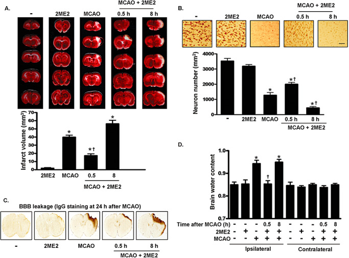
MCAO‐induced brain damage is regulated by 2ME2. A. Infarct area evaluated by TTC staining. Representative sections from the indicated treatments after 4 days of recovery are shown. The infarct area was measured in 12 sequential sections. B. Quantification of NeuN‐positive cells using immunohistochemistry after the indicated treatments. Brain sections were obtained 4 days later and immunostained with anti‐NeuN antibody. Scale bar, 100 µm. C. IgG staining in sections of the rat brain of sham‐control, MCAO and MCAO plus 2ME2 groups, respectively. Brain sections were obtained 24 h later and immunostained with goat anti‐rat IgG biotin. D. The hemispheres were separated and weighed immediately after removal and again after drying in an oven at 105°C for 24 h. In panels A, B and D, data are presented as mean (SD) (n = 4). Statistical analysis was carried out using the one‐way ANOVA with appropriate post hoc tests; *P < 0.05 vs. the sham‐control group. †P < 0.05 vs. MCAO group. Abbreviations: MCAO = middle cerebral artery occlusion; 2ME2 = 2‐methoxyestradiol; TTC = 2,3,5‐triphenyltetrazolium chloride; NeuN = Neuronal Nuclei; IgG = immunoglobulin; SD = standard deviation; BBB = blood‐brain barrier.
