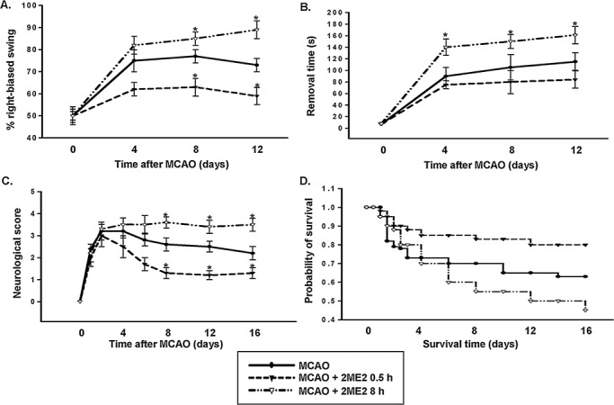
Figure 3.

Role of early‐ and late‐phase HIF‐1αexpression in ischemic brain injury. A–C. Line graphs show temporal profiles of functional recovery from rats in each treatment. The distinct effects of rats that received 2ME2 0.5 h or 8 h after subjected to MCAO were detected using the swing test (A), adhesive‐removal patch test (B) and scoring of neurological deficit (C). D. Kaplan–Meier survival analysis after MCAO in the indicated 2ME2 treatment. In panels A–C, statistical analysis was carried out using the one‐way ANOVA with appropriate post hoc tests; *P < 0.05 vs. MCAO group. HIF‐1α = hypoxia‐inducible factor‐1‐alpha; MCAO = middle cerebral artery occlusion; 2ME2 = 2‐methoxyestradiol.
