
Figure 2.
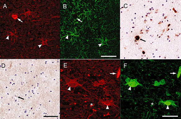
TG2 immunoreactivity in astrocytes. Double immunofluorescence staining showed the presence of TG2 immunoreactivity (A) in GFAP positive astrocytes (B). Scale bar: 40 µm. TG2 immunoreactivity in astrocytes (C) and endothelium is abolished after preadsorption of the anti‐TG2 antiserum with guinea pig TG2 (D). Scale bar: 100 µm. TG2 immunoreactivity in astrocytes, endothelium and extracellularly (D) co‐localized with Fn, an ECM protein (F). Scale bar: 40 µm. Arrow: blood vessel, arrowhead: astrocyte, asterisk: extracellular.