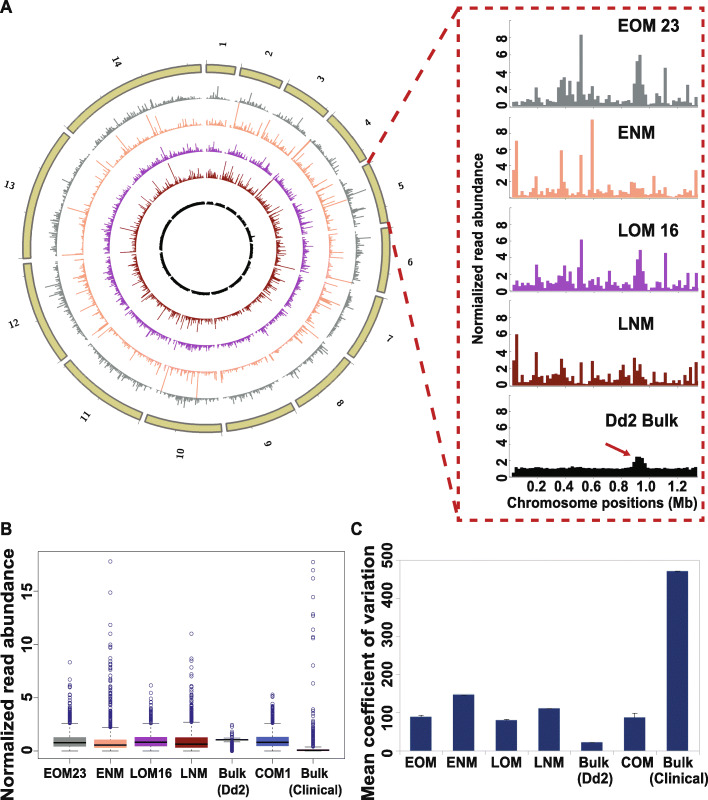
Fig. 3.
Samples amplified by optimized MALBAC display improved uniformity of read abundance. a Normalized read abundance across the genome. The reference genome was divided into 20-kb bins and read counts in each bin were normalized by the mean read count in each sample. The circles of the plot represent (from outside to inside): chromosomes 1 to 14 (tan); one EOM sample (#23, grey); one ENM sample (#3, orange); one LOM sample (#16, purple); one LNM sample (#2, dark red); Dd2 bulk genomic DNA (black). The zoomed panel shows the read distribution across chromosome 5, which contains a known CNV (Pfmdr1 amplification, arrow on Dd2 bulk sample). b Distribution of normalized read abundance values for all bins. The boxes were drawn from Q1 (25th percentiles) to Q3 (75th percentiles) with a horizontal line drawn in the middle to denote the median of normalized read abundance for each sample. Outliers, above the highest point of the upper whisker (Q3+ 1.5 × IQR) or below the lowest point of the lower whisker (Q1−1.5 × IQR), are depicted with circles. One sample from each type is represented (see all samples in Additional file 1: Figure S3C). c Coefficient of variation of normalized read abundance. The average and SD (error bars) coefficient of variation for all samples from each type is represented (EOM: 13 samples; ENM: 1 sample; LOM: 10 samples; LNM: 1 sample; Dd2 bulk: 1 sample; COM: 2 samples; Clinical bulk: 1 sample). See “Methods” for calculation

