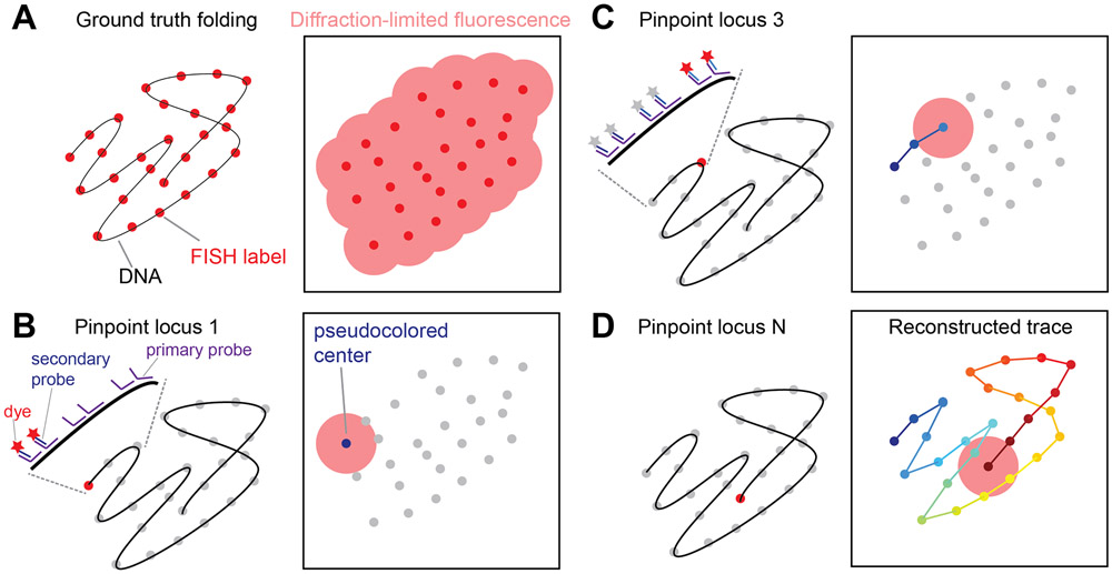
Figure 1.
Concept of Chromatin Tracing. (A) Conventional DNA fluorescence in situ hybridization (FISH) cannot distinguish and resolve many genomic loci along the same chromatin. (B, C) In chromatin tracing, primary FISH probes are hybridized to all genomic loci of interest. Then dye-labeled secondary FISH probes help sequentially visualize the genomic loci as individual diffraction-limited spots, the centers of which can be pinpointed with nanoscale accuracy. The procedures to pinpoint locus 1 (B) and locus 3 (C) are shown. (D) After all loci are visualized and pinpointed, their center positions are linked according to their genomic identities to reconstruct the chromatin trace.

