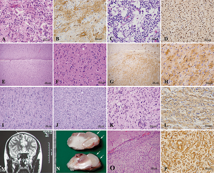
Figure 3.

(A–L) Histopathological findings of mixed neuronal–glial tumors. Gangliogliomas are composed of large ganglion‐like cells and neoplastic glial cells (A), and show a bushy pattern of CD34 positivity (B). Dysembryoplastic neuroepithelial tumors with a microcystic morphology contain isolated, mature large neurons (C) and oligodendroglia‐like cells, which are immunolabeled with Olig2 antibody (D). (E–H) Mixed neuronal–glial tumors with preliminary features of ganglioglioma. With low magnification, obvious cortical architectural disturbances can be seen (E). Note a normal appearance of cortex seen in the upper part. High magnification shows randomly scattered neurons and glial cells of somewhat high cellularity (F). CD34 immunostainig reveals diffuse and solitary patterns (G,H). (I,J) Mixed neuronal–glial tumors with preliminary features of dysembryoplastic neuroepithelial tumors. The area with normal neurons surrounded by proliferating oligodendrocytes (I) is adjacent to the area with an admixture of neuronal and proliferating oligodendrocytic elements (J). (K,L) Pleomorphic xanthoastrocytoma shows nuclear pleomorphism and xanthomatous change (K), as well as diffuse CD34 immunostaining (L). (M–P) Neuroimaging and histopathological findings of meningioangiomatosis. T2‐weighted magnetic resonance imaging (MRI) image displays an abnormal signal in the cortex of the right frontal lobe (M, arrow). Macroscopically, partial gyri appear tumid and pale (N, arrows). Microscopically, there were proliferating spindle cells and vessels invading into the superficial cortex with rich collagen deposition (O,P). (A,C,E,F,I,J,K,O) hematoxylin and eosin (H&E); (B,G,H,L) CD34 immunostaining; (D) Olig2 immunostaining; (P) vimentin immunostaining.
