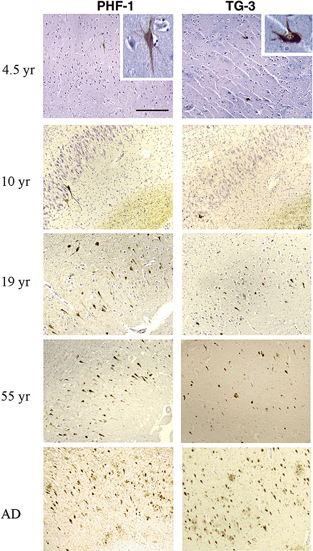
Figure 1.

Progression of NFT formation with age in NPC. Paraffin‐embedded sections from hippocampus (for the 6‐year‐old case only the temporal cortex was available) were immunostained with the PHF‐1 and TG‐3 antibodies (brown), and each section was counter‐stained with Hematoxylin to visualize the nuclei (violet) of all cells in a given field. Light micrographs from NPC cases with different ages show the relative numbers and distribution of NFT in CA1 region of hippocampus, which look similar to those in a typical AD case (20×, scale bar = 80 microns).
