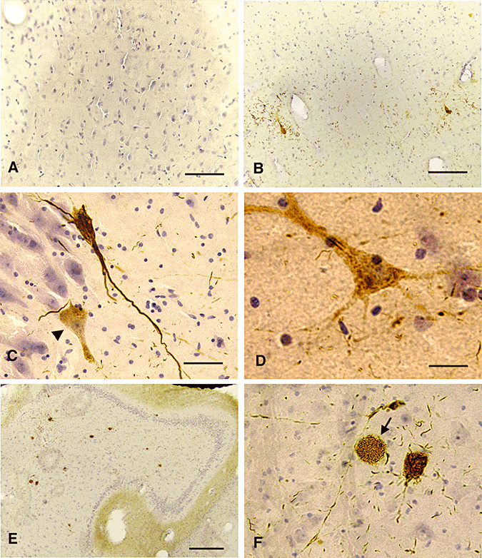
Figure 2.

NFT pathology in juvenile NPC. Sections from the hippocampus of the 7‐month‐old (A), the 4.5‐year‐old (B) and an 10‐year‐old NPC cases (C–F) were stained with PHF‐1 (C–D) or TG‐3 (A, B, E, F) and counter‐stained with hematoxylin (A, B, F, bar = 80 microns; C, D, bar = 40 microns; E, bar = 160 microns). PHF‐1 or TG‐3 did not stain the neurons in the 7‐month‐old NPC case (A). A mature NFT was developed in the CA1 region of the 4.5‐year‐old NPC case (B). Similarly, fully‐developed NFT were stained with PHF‐1 in the 10‐year‐old NPC case (C) and some neurons with diffuse PHF‐1 immunoreactivity were seen (arrowhead in C, and D). Increased numbers of NFT were common in the CA4 hippocampal region in the 10‐year‐old NPC case (E), and some neurons with only lipid storage were also noted (F).
