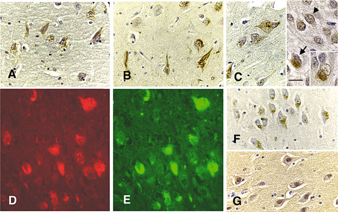
Figure 5.

Mitotic epitopes demarcating NFT. Hippocampal sections from a 31‐year‐old case were immunostained with MPM‐2 (panels A, D), H14 (panel B), H5 (panels C, E), cdc2 (panel F) and cyclin B1 (G) and counter‐stained with hematoxylin (all panels, bar = 40 microns). Classic NFT were stained with every antibody (panels A and D for MPM‐2, and panel B for H14, and C and E, H‐5), but cdc2 and cyclin B1 staining of some neurons lacking obvious NFT was more prominent (panels F and G, respectively). The H5 staining shows a shift in distribution of the enzyme from the nucleus (panel C, arrowhead) to the cytoplasm (panel C, arrow, and inset). MPM‐2 (panel D, red) and H5 (panel E, green) colocalize in the same NFT.
