Figure 2a.

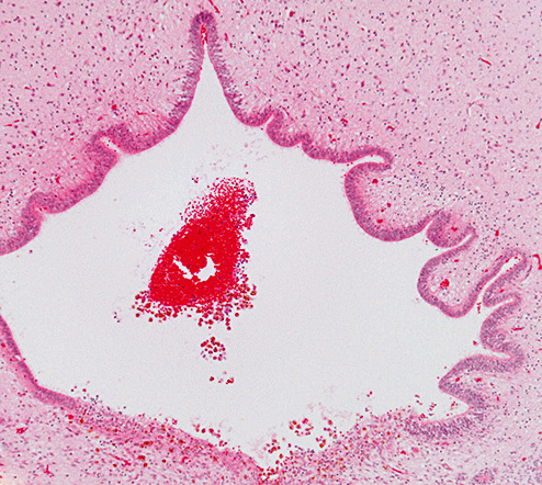
Photomicrograph showing a small blood clot in the proximal aqueduct of Case 1. The posterior aspect (lower part of figure) exhibits hemosiderin‐containing macrophages within the tissue (hematoxylin and eosin stain; magnification 100×).

Photomicrograph showing a small blood clot in the proximal aqueduct of Case 1. The posterior aspect (lower part of figure) exhibits hemosiderin‐containing macrophages within the tissue (hematoxylin and eosin stain; magnification 100×).