
Figure 2.
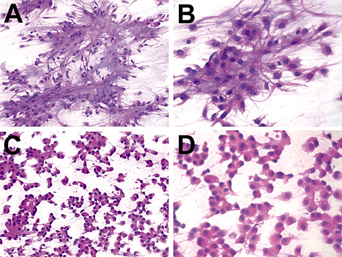
Intraoperative preparation of chordoid glioma. The intraoperative smears on case 1 showed a biphasic neoplasm. Some areas contained clumps of cells with abundant pink cytoplasm and long fibrillar processes in a myxoid background (A & B). Other areas had cohesive clusters of epithelioid cells with abundant eosinophilic cytoplasm and shorter delicate processes without a myxoid background (C & D). Both types of cells had medium‐sized nuclei with 1–2 small nucleoli. No mitoses, necrosis, or bizarre cellular atypia were observed.