
Figure 4.
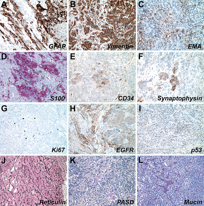
Immunohistochemical and histochemical profile of chordoid glioma. Case 1 tumor cells were positive for GFAP (A) and vimentin (B), and showed focal positivity for EMA (C), S100 (D), CD34 (E), and synaptophysin (F). Ki67 proliferation index was low at ∼3% (G). Some areas of the tumor expressed EGFR (H). It did not show a clonal pattern of p53 immunoreactivity (I). The tumor had an extensive reticulin network (J) and stroma with some mucopolysaccharides but no mucin (K and L). Abbreviations: GFAP = glial fibrillary acidic protein; EMA = epithelial membrane antigen; EGFR = epidermal growth factor receptor; PASD = Periodic Acid‐Schiff with diastase.