Figure 1.

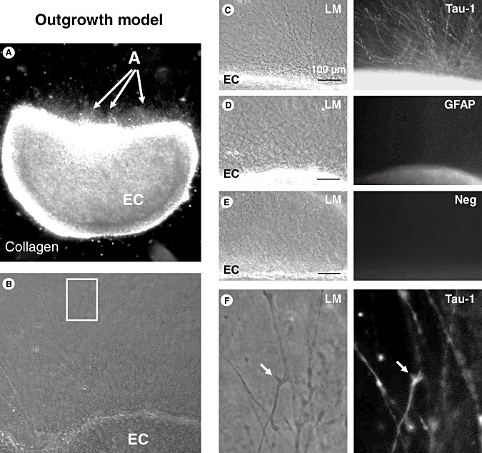
Outgrowth model. A. Outgrowth assay (see Material and Methods section for details); the entorhinal cortex (EC) explant was embedded in a three‐dimensional collagen I gel matrix; the density of the outgrowing neurites (arrows) was photo‐documented at higher magnification and quantified by two blinded investigators. B. Neurite outgrowth in control organotypic brain slices. The white box indicates the area shown at higher magnification in 1F. C. Multiple neurites are visible by light‐microscopy (LM) growing out of the concave part of the EC explant. Immunofluorescence staining using specific antibodies reveals immunreactivity against Tau‐1, which is found on neurites but not on glial cells. D. No immunoreactivity of the extensions is detectable using a specific antibody against glial fibrillary acidic protein (GFAP) as a marker for astrocytes and their extensions. E. No immunoreactivity of the extensions is found using the secondary antibody alone as a negative control. F. Higher magnification of the white box in 1B: Tau‐1‐labeled neurites at light microscopy (LM) show characteristic growth cones that are also immunoreactive for Tau‐1 as a marker for neurites.
