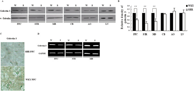
Figure 2.

Comparison of galectin‐3 expression between ADHD and WKY rats. A. Galectin‐3 protein was analyzed by Western blot in prefrontal cortex (PFC), striatum (STR), midbrain (MB), cerebellum (CB), aorta (AO), liver (LV); B. Gray density analysis of the bands in A. The bands on X‐ray films was scanned and analyzed by the software Image J (Wayne Rasband, National Institute of Health, USA). Data were given as an average of relative density with standard errors, where n = 5. **P < 0.01; C. Immunostaining of the PFC slice was with galectin‐3 antibody, and cells were viewed in 400× magnifications. PFC slices of the brain were incubated with rabbit anti‐galectin‐3 antibody, HRP‐conjugated goat anti‐rabbit immunoglobulin and DAB sequentially, and counterstained with methyl green. The image was the representative of three independent experiments. D. Analysis of galectin‐3 mRNA level in PFC, STR and MB by RT–PCR. Abbreviations: ADHD = attention deficit hyperactivity disorder; DAB = diaminobenzidine tetrahydrochloride; HRP = horseradish peroxidase; RT–PCR = reverse transcriptase–polymerase chain reaction; SHR(S) = spontaneously hypertensive rats; WKY(W) = Wistar–Kyoto rats.
