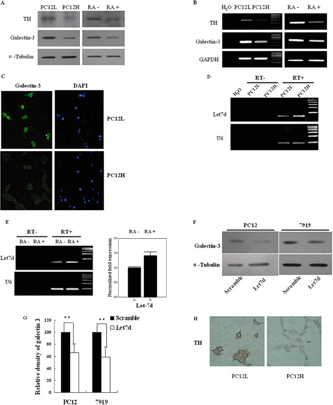
Figure 5.

Galectin‐3, TH and rno‐let‐7d expressions in PC12 cells. A. Galectin‐3 and TH protein levels were detected by Western blot. Galectin‐3 and TH expressions were analyzed in both PC12L and PC12H cells at 48 h after passage, which showed that the expressions were higher in PC12L cells than in PC12H cells. Galectin‐3 and TH expressions in PC12L cells were then investigated at 6 days after incubation with 1 µmol/L RA. B. Galectin‐3 and TH mRNA levels were simultaneously detected by RT–PCR. C. Immunofluorescence staining of galectin‐3 in PC12L and PC12H cells, in 200× magnifications. D. Mature rno‐let‐7d was detected in PC12L and PC12H cells. Reactions without reverse transcriptase (RT‐) were as a negative control. E. Mature rno‐let‐7d was detected by either stem‐loop RT‐PCR (left panel) or real‐time PCR (right panel) in PC12L cells before and after 48 h incubation with 1 µmol/L RA. Reaction without reverse transcriptase (RT‐) was used as a negative control. The results were the representative of three independent experiments. F. In PC12L cells and CBRH‐7919 cells, galectin‐3 protein expression was reduced after transient transfection with rno‐let‐7d precursor. G. Density analysis of Western blot results in F. Density of the bands on X‐ray films was scanned, quantified by Image J (Wayne Rasband, National Institute of Health, USA). Data were shown as an average of relative density with standard errors (**P < 0.01), where n = 5. H. Immunohistochemistry observation of TH in PC12L and PC12H cells. Magnification: 200×. Abbreviations: DAPI = 4′,6‐diamidino‐2‐phenylindole; PC12L = low differentiated pheochromocytoma cells; PC12H = high differentiated pheochromocytoma PC12 cells; RA = retinoic acid; RT–PCR = reverse transcriptase–polymerase chain reaction; TH = tyrosine hydroxylase.
